
SOFTWARE
Intelligent Imaging.
Precise Decisions.
Advanced software solutions designed to unlock the full potential of digital dental imaging. Intuitive, powerful and scalable platforms created to optimize clinical workflows and enhance diagnostic accuracy.
DEEP-VIEW
Intraoral Imaging Software


Key Capabilities
Integrated Multi-System Imaging Platform
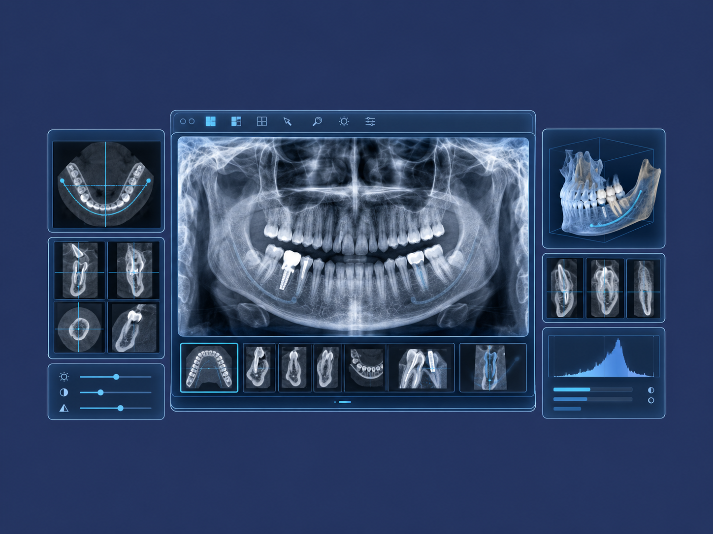
Deep-View centralizes image acquisition from multiple devices—including intraoral sensors, panoramic systems, cameras and scanners—providing a unified, consistent interface for managing all clinical data.
High-Precision Image Processing
Advanced algorithms optimize image quality through mathematical filters—including contrast, brightness, edge enhancement and pseudo-3D—facilitating detailed analysis of anatomical structures, even under low-contrast conditions.
Advanced Visualization and Multidimensional Analysis
Deep-View enables data exploration in multiple formats, including panoramic, axial and cross-sectional views, 3D visualization and volumetric reconstructions. Advanced measurement tools—including density assessment and Hounsfield-type scales—support in-depth clinical analysis.
Advanced DICOM Data Management
All images are organized, archived and kept accessible within a DICOM-compliant system, ensuring traceability, proper organization and continuity of the patient record.
Optimized Cross-Platform User Experience
An intuitive interface designed to reduce operational steps and improve clinical efficiency, available on Windows, macOS, iOS and Android for maximum flexibility.
Guided Workflow and Operational Reliability
An integrated system that identifies anomalies, guides the user and supports safe, continuous operation, reducing errors and optimizing overall performance.
CORE FUNCTIONS
Integration with multiple devices (TWAIN)
Multiple database management
Real-time image processing
Advanced filters and precise analysis
Customizable templates (FMX)
Export in DICOM and other compatible standard formats
DEEP-VIEW SEAMLESSLY INTEGRATES WITH:
X-VIEW 2D, X-VIEW 3D, I-View Silver & I-View Gold
XELIS – Advanced Implant Planning
Xelis is an advanced implant planning software solution designed to maximize the potential of CBCT data and support clinicians throughout every stage of treatment.
Developed to provide a precise and comprehensive view of the patient’s anatomy, it enables clinicians to simulate implant positioning, assess bone structures and identify critical areas with a high degree of reliability.
With its powerful 2D/3D visualization, analysis and measurement tools, Xelis supports clinical decision-making while enhancing procedural safety and predictability.
Designed for modern implantology, Xelis optimizes preoperative planning and integrates seamlessly into a complete digital workflow.
Key Capabilities
3D Implant Simulation
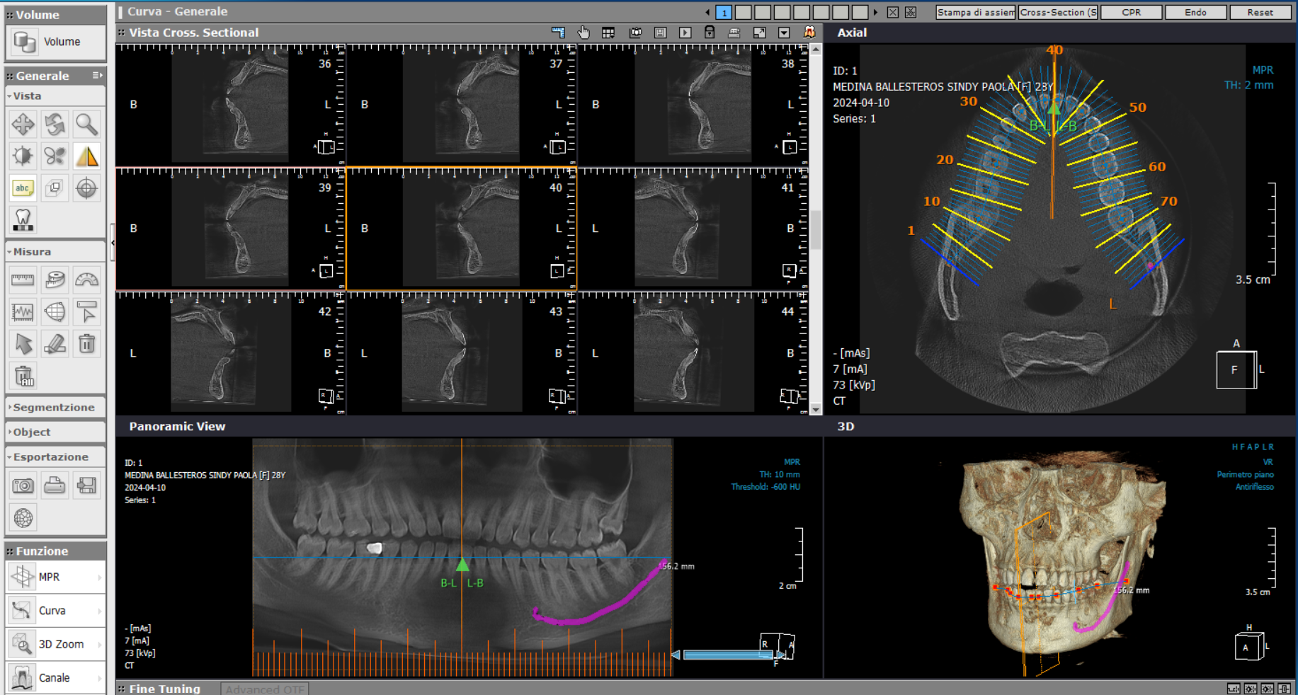
Virtually position implants within the patient’s CBCT volume and adjust their diameter, length, depth, angulation and insertion axis according to the available bone and the planned surgical approach.
MPR and Cross-Sectional Analysis
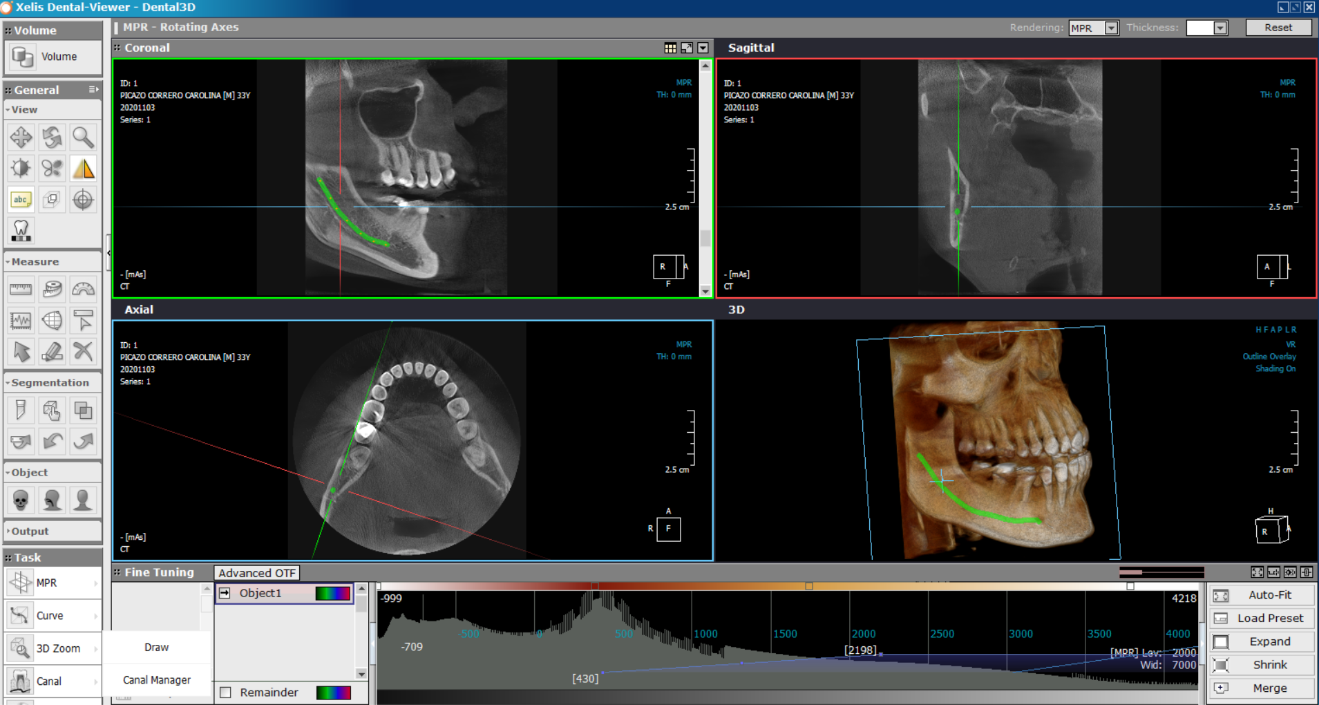
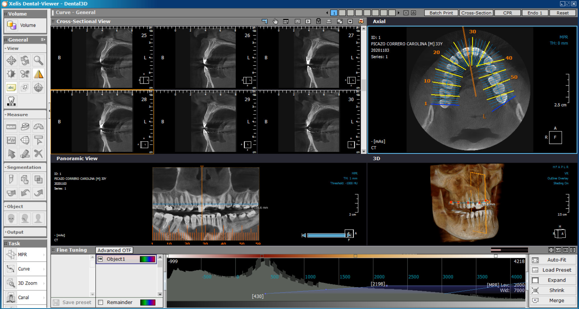
Navigate through axial, coronal, sagittal, panoramic and cross-sectional views. Reconstruct the dental arch and evaluate bone morphology, available height and width, and relationships with adjacent anatomical structures.
DICOM-Based CBCT Visualization
Import and review CBCT datasets in a DICOM environment, with direct access to the complete volumetric examination and the anatomical information required for implant planning.
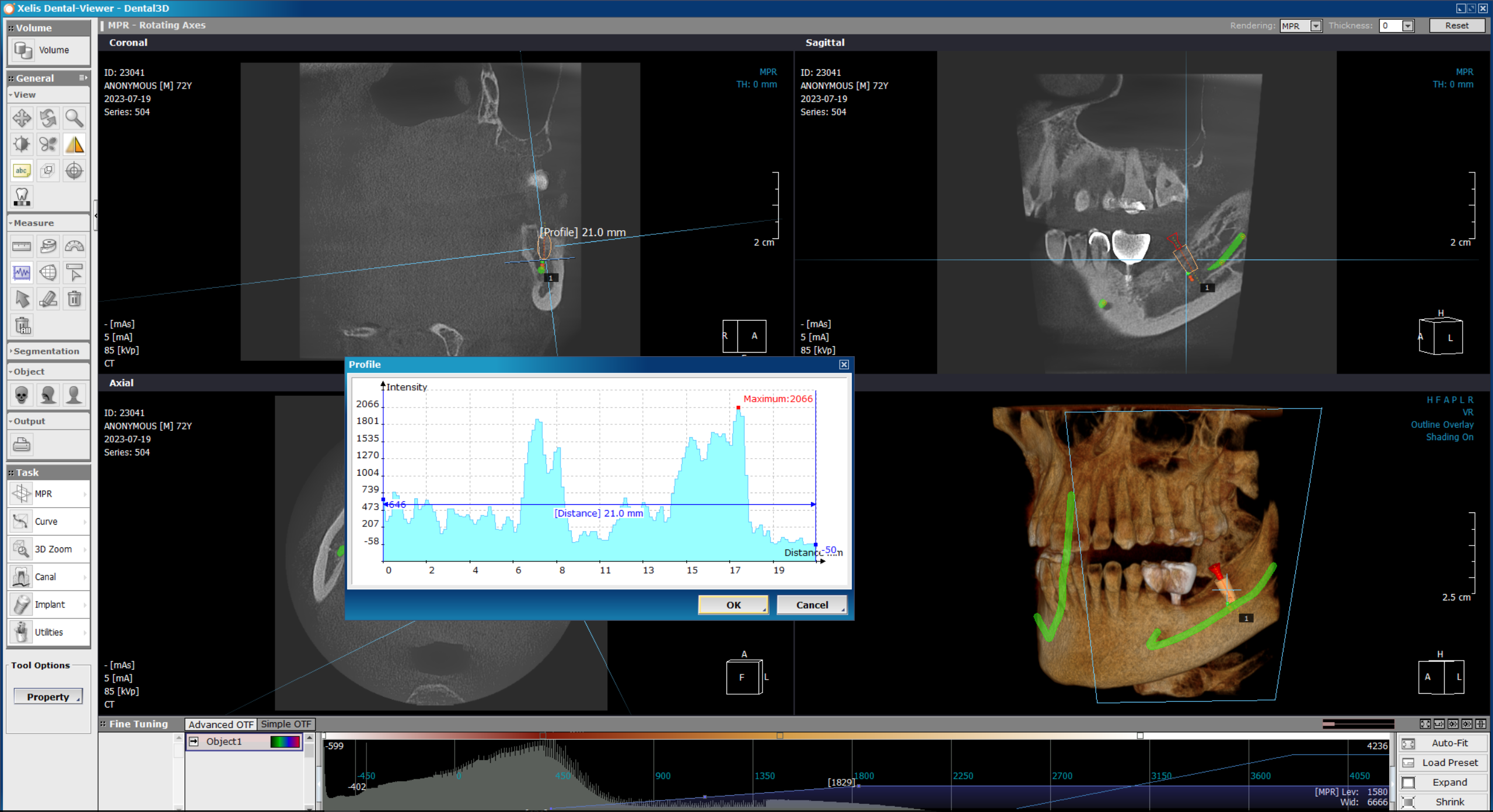
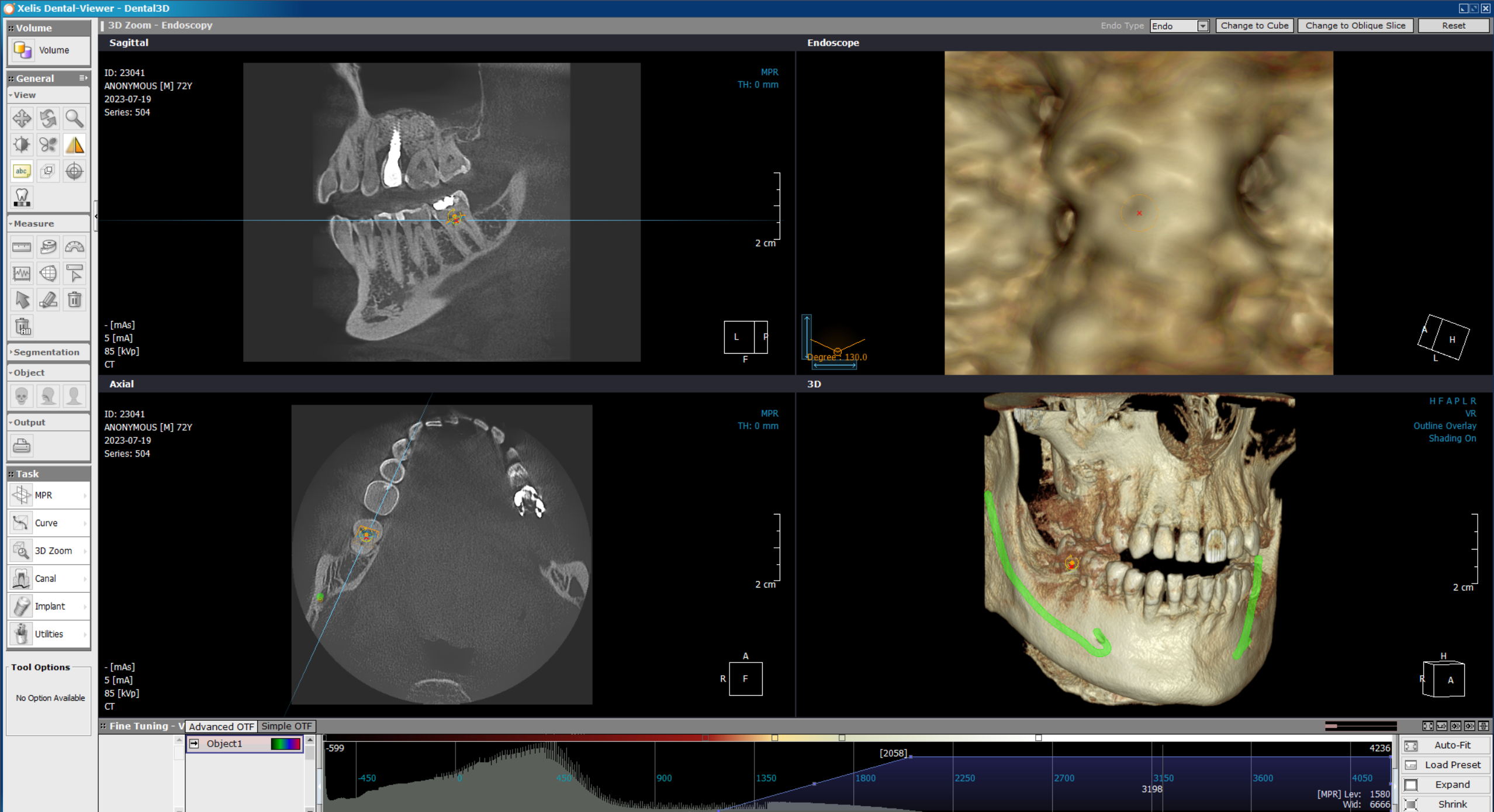
Mandibular Canal Tracing
Trace and visualize the mandibular canal throughout the planning process. Review the implant’s position and distance in relation to the inferior alveolar nerve and other critical anatomical structures.
Bone Assessment and Implant Library
Use measurement and bone-density assessment tools to evaluate the proposed implant site. Select the appropriate implant from an integrated library containing systems and dimensions from leading manufacturers.
STL Export for Digital Surgical Workflows
The Advanced version enables STL export for integration with surgical-guide design, 3D manufacturing and other digital planning workflows, connecting CBCT-based implant planning with the subsequent surgical process.
CORE FUNCTIONS
Integrated Xelis DBM patient and case database
Multi-user network environment for up to 10 users
Case storage, retrieval and structured data management
Customizable toolbars and viewing layouts
Bookmarking and annotation tools
Customizable templates and implant planning reports
Image and case export to CD or USB
Batch printing of axial, panoramic and cross-sectional views
Dynamic Lightbox for comparative image review
Image stitching and reconstruction tools
DICOM printing and media creation
PACS integration and clinical data exchange
A SOLUTION FOR EVERY CLINICAL NEED
Xelis is available in two versions—ESSENTIAL and ADVANCED—designed to meet different clinical requirements and support the digital workflows of modern dental practices.
Each version provides a dedicated set of features, allowing clinicians to select the solution that best suits their practice—from essential implant planning to advanced guided-surgery workflows.
| FEATURES | ESSENTIAL | ADVANCED |
|---|---|---|
| Xelis DBM Database | ✓ | ✓ |
| 3D Tools (MPR, Cross-Sections, Measurements) | ✓ | ✓ |
| Implant Simulation | ✓ | ✓ |
| Mandibular Canal Tracing | ✓ | ✓ |
| Image Export (CD / USB) | ✓ | ✓ |
| Multi-User Access (Up to 10 Users) | ✓ | ✓ |
| ADVANCED FEATURES | ||
| STL Export | — | ✓ |
| Batch Printing | — | ✓ |
| Dynamic Lightbox (DLB) | — | ✓ |
| Image Fusion / Reconstruction | — | ✓ |
| Advanced Reporting and Case Management | — | ✓ |
| DICOM Printing and CD Burning | — | ✓ |
XELIS SEAMLESSLY INTEGRATES WITH:
X-VIEW 3D, X-VIEW 3D PAN, X-VIEW 3D PAN CEPH
AUDAXCEPH – Orthodontic, Cephalometric Analysis & Treatment Simulation Suite
AudaxCeph is a specialized software solution with the essential tools for orthodontic assessment and treatment design together in a single, efficient digital environment.
Designed to streamline the diagnostic workflow, it enables complete analyses to be performed in seconds using automated tools and predefined cephalometric models.
With VTO simulation, image superimposition and growth projection capabilities, AudaxCeph provides a comprehensive view of treatment evolution, supporting clinical decision-making and facilitating clear communication with the patient.
Key Capabilities
Fast, Automated Cephalometric Analysis
AudaxCeph performs complete cephalometric analyses in seconds using predefined models and an optimized workflow, significantly reducing the time required for clinical assessment.
Advanced Superimposition and Comparative Analysis
AudaxCeph enables multiple analyses and images to be superimposed using point-based, structural and rotational methods, facilitating the evaluation of treatment progress over time.
AI-Powered Anatomical Landmark Detection – TINA
The system automatically identifies anatomical landmarks on radiographic images, reducing manual steps while supporting consistent, accurate and reproducible analyses.
Growth Projection and Long-Term Planning
Integrated growth-projection tools (VTG) help clinicians evaluate potential skeletal development and plan appropriate long-term treatment strategies.

Advanced Treatment Simulation – VTO
Clinicians can simulate different orthodontic and surgical treatment scenarios by incorporating the patient’s anatomical parameters into detailed visual treatment projections.
Automated Reports and Clinical Documentation
Analysis results can be automatically generated in PDF, vector and Excel formats, simplifying clinical documentation, case monitoring and communication with patients and colleagues.
Intuitive Interface and Optimized Workflow
A modern interface with interactive tools—including drag-and-drop functionality and automated processes—streamlines daily operations and reduces the learning curve.
Patient Image and Case Management
AudaxCeph organizes images, documents and clinical cases within a structured, easily accessible environment designed to support modern orthodontic workflows.
CORE FUNCTIONS
Structured patient and case data management
Customizable cephalometric analysis templates
Quick review and manual adjustment of anatomical landmarks
Seamless navigation between analyses, images and superimpositions
Longitudinal comparison across different treatment stages
Automated report generation in PDF, vector and Excel formats
Case data export and sharing
Integration with leading Practice Management Systems (PMS) for a connected clinical workflow
AUDAXCEPH WORKS SEAMLESSLY WITH:
X-VIEW 2D PAN CEPH, X-VIEW 3D PAN CEPH
DFO – Digital Dentofacial Analysis
The optional DFO (Digital Facial Orthopedics) module provides clinicians with dedicated tools for evaluating skeletal structures and soft-tissue profiles using digital images.
Through an optimized workflow and automated functions, DFO enables fast, consistent and reproducible analyses, supporting diagnostic assessment and improving efficiency in everyday clinical practice.
Key Capabilities
Comprehensive Dentofacial Analysis from Digital Images
DFO combines dental and facial images to support detailed cephalometric and morphological assessments of skeletal structures and soft-tissue profiles.
Integration with the Patient’s Clinical Record
Analysis results can be incorporated directly into the patient’s clinical documentation, facilitating case management and longitudinal follow-up.
Streamlined Analysis Workflow
DFO simplifies the different stages of dentofacial analysis compared with traditional methods, reducing processing time and minimizing manual operations.


Automated Measurements and Reproducible Results
Once the anatomical landmarks have been defined, the system automatically calculates angles, distances and proportions, supporting consistent and reproducible evaluations.
Customizable Clinical Analysis Protocols
Clinicians can create or modify analysis protocols according to their clinical requirements, preferred methodology and individual case needs.
Clinical Data Export and Collaboration
Analyses and results can be exported and shared with colleagues, referring clinicians and external specialists for efficient interdisciplinary collaboration.
CORE FUNCTIONS
Import and management of dental and facial images
Anatomical landmark placement, review and manual adjustment
Automatic recalculation after landmark modifications
Creation and customization of clinical analysis protocols
Structured visualization of measurements, angles and proportions
Longitudinal comparison of analyses throughout treatment
Patient case organization and clinical documentation
Export and sharing of analysis results