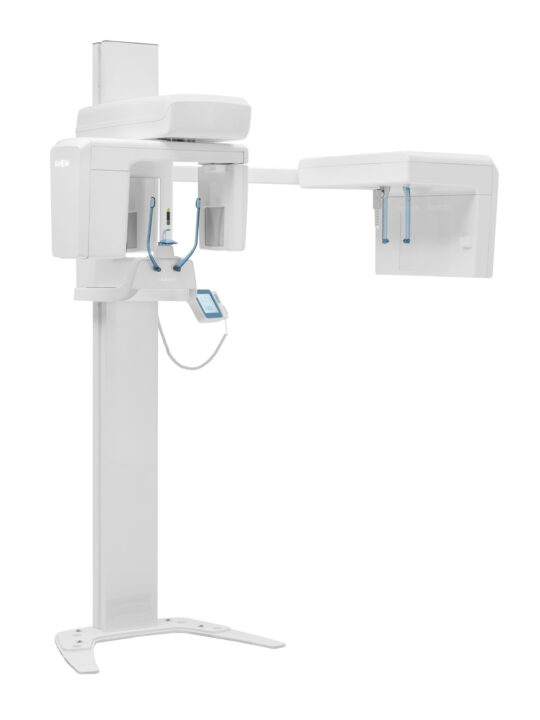
X-VIEW 2D PAN

Elegant Design.

Outstanding Imaging.

More than a panoramic imaging system, X-VIEW 2D PAN is an imaging platform designed to evolve with your practice.

Its advanced imaging technology, outstanding image quality, and intuitive workflow deliver a precise, efficient clinical experience, ready to meet the demands of modern dentistry.

ONE PLATFORM. THREE CONFIGURATIONS.

Available in three configurations, from a high-performance panoramic unit to a fully integrated cephalometric solution, X-VIEW 2D PAN provides the flexibility to meet your clinical needs while protecting your investment for the future.

2D PAN BASIC2D PAN UP2D PAN CEPH
TAKE PANORAMIC IMAGING FURTHER WITH DEEP‑AI

Discover how Deep‑AI, seamlessly integrated into Deep‑View, brings out the full potential of panoramic images acquired with X‑VIEW 2D PAN—enhancing clarity, contrast, sharpness, and image consistency while preserving the original anatomical information.

Available as an optional software module.

EXPLORE DEEP-AI
FEATURES

Remarkable features that
cannot be seen:

THE FULL POTENTIAL OF X‑VIEW 2D PAN IN AN INTEGRATED DIGITAL ENVIRONMENT

The X-VIEW 2D PAN is supported by a comprehensive software ecosystem designed to simplify every stage of the imaging workflow, from image acquisition and management to advanced analysis, treatment planning, and patient communication.

Working seamlessly together, Trident software solutions help clinicians capture, organize, analyze, and present diagnostic information with efficiency and confidence.

The ecosystem includes:

Deep-View

The imaging platform for acquisition, visualization, storage, and management of 2D dental images.


AudaxCeph

Advanced cephalometric analysis and comprehensive orthodontic treatment planning.


DFO

Treatment simulation and visual communication tools designed to improve patient understanding and engagement.

Together, they extend the capabilities of the X-VIEW 2D PAN, supporting clinicians from image acquisition to diagnosis, treatment planning, and patient communication.

DEEP-VIEW

Deep‑View is the core of Trident’s digital imaging ecosystem, a platform developed to acquire, visualize, and manage dental radiographic images within a single working environment.

Compatible with intraoral, panoramic, and cephalometric imaging systems, it combines advanced image visualization and optimization tools with a clear and intuitive interface. This simplifies daily activities and provides quick access to images and patient clinical information.

From image acquisition and processing to the organization of examinations and patient data, Deep‑View connects every stage of the workflow, delivering a faster, more organized, and efficient experience.

FEATURE HIGHLIGHTS

  • Fast image acquisition and visualization
  • Intuitive patient and examination management
  • Advanced image visualization tools
  • Image enhancement and optimization functions
  • DICOM connectivity
  • Compatibility with intraoral, panoramic, and cephalometric imaging systems

AUDAXCEPH: Orthodontic Cephalometric Analysis & Treatment Simulation Suite

AudaxCeph Ultimate is professional software that redefines orthodontic treatment planning.

It provides precise, automated cephalometric analyses, intuitive VTO simulations, and reliable growth predictions. Its intelligent design streamlines the analysis process and supports confident clinical decision-making, delivering a faster, more precise, and efficient workflow.

FEATURE HIGHLIGHTS

  • Automatic detection of anatomical landmarks
  • Automated cephalometric tracing and analysis
  • Visual Treatment Objective (VTO) simulation
  • Craniofacial growth prediction
  • Image superimposition to evaluate changes
  • Treatment planning and simulation tools
  • Clear and customizable results

DFO: Dental - Facial and Orthodontics Analysis

DFO is a dedicated software solution for orthodontic tracing and cephalometric analysis.

The user manually identifies the required anatomical landmarks, and the software automatically calculates the measurements and generates the complete tracing on screen. Its clear and practical workflow simplifies the analysis process and supports efficient case evaluation and documentation.

FEATURE HIGHLIGHTS

  • Manual identification of anatomical landmarks
  • Automatic tracing generation based on the selected landmarks
  • Automatic calculation of cephalometric measurements and results
  • Differential and orthodontic analysis
  • Layer-based analysis and visualization
  • Interactive tutor to guide the analysis process
  • DICOM image storage and management

Correct patient positioning is essential for high-quality panoramic and cephalometric imaging. X‑VIEW 2D PAN combines intuitive positioning aids that simplify patient alignment, improve examination reproducibility, and help reduce positioning errors and retakes—supporting consistent image quality from the very first acquisition.

PRECISE POSITIONING, MADE SIMPLE


ACCURATE ALIGNMENT
Dual laser guides ensure accurate alignment of the Median Sagittal and Frankfurt planes, while the integrated mirror helps patients position themselves correctly.

STABLE AND REPRODUCIBLE
The adjustable nasion support, temple supports, and ergonomic bite block work together to stabilize the patient’s head and dentition throughout image acquisition.

COMFORTABLE AND ACCESSIBLE
Motorized height adjustment and an open design simplify positioning for patients of different heights, including wheelchair users, while providing easy access for the operator.

Together, these features make patient positioning faster and more repeatable, helping reduce errors and retakes while supporting consistent image quality.

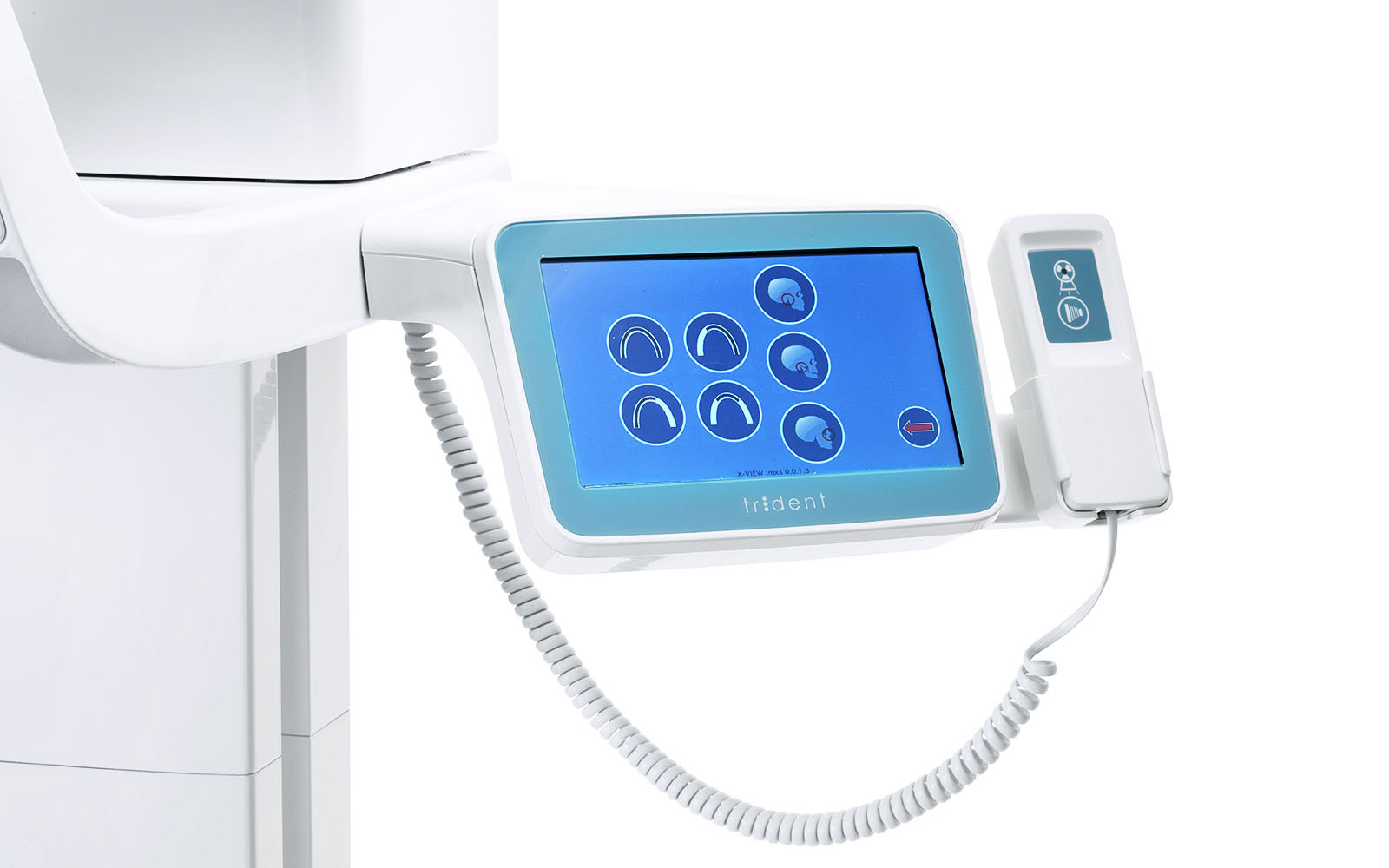
The 7-inch LCD touchscreen integrates advanced technology to provide fast and intuitive control of the unit. Programs, parameters, and functions are easily accessible through touch controls within a clear, well-organized interface that simplifies every operation.

DIRECT CONTROL
The touchscreen interface allows operators to select examination programs and configure exposure parameters directly from the unit.

INTUITIVE NAVIGATION
The logical menu structure simplifies every selection and keeps relevant information clearly visible throughout examination setup.

UNIVERSAL ICONS
A clear visual language makes functions, parameters, and available options immediately recognizable, facilitating direct interaction with the unit.

MULTILINGUAL INTERFACE
Instructions available in multiple languages guide the operator step by step, supporting smooth and consistent use across different clinical environments.

X‑VIEW 2D PAN:

The three models in detail

X-VIEW 2D PAN BASIC

Everything you need to bring panoramic imaging into your practice.

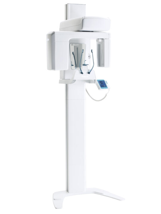
X‑VIEW 2D PAN BASIC is the ideal choice for dental practices seeking a reliable, intuitive and cost-effective panoramic imaging solution. It combines outstanding image quality with a streamlined workflow, helping clinicians perform routine examinations efficiently and with confidence.

Easy to install, operate and maintain, X‑VIEW 2D PAN BASIC delivers all the essential panoramic capabilities in a compact and accessible system, without unnecessary complexity. A smart investment for practices entering the world of extraoral imaging or looking for a dependable unit dedicated to panoramic examinations.


HIGH-RESOLUTION CMOS SENSOR FOR PANORAMIC IMAGING

  • 30 × 15 cm adult panoramic examination in 15.5 s
  • 30 × 13 cm child panoramic examination in 14.8 s

DEEP‑VIEW + DEEP‑AI

X‑VIEW 2D PAN BASIC integrates seamlessly with Deep‑View, creating a smooth digital workflow from image acquisition to visualization and management.

When combined with the optional Deep‑AI module, artificial intelligence automatically enhances panoramic images by optimizing clarity, contrast, sharpness and image consistency—while preserving the original anatomical information.

Essential Features. Outstanding Performance

  • High-quality panoramic imaging for everyday clinical needs
  • Optional AI-powered image enhancement with Deep‑AI
  • Complete selection of standard examination programs
  • Fast and intuitive workflow
  • Precise and comfortable patient positioning
  • Open and accessible design
  • Reliable performance and straightforward maintenance

X-VIEW 2D PAN UP

Start with Panoramic.

Expand when you’re ready.

X‑VIEW 2D PAN UP offers the same panoramic imaging performance, intuitive workflow and everyday reliability as X‑VIEW 2D PAN BASIC, with one important advantage: it is designed from the outset for future cephalometric expansion.

The unit incorporates the mechanical and user-interface provisions required for a subsequent upgrade to X‑VIEW 2D PAN CEPH. When the practice is ready, the system can be expanded by adding the cephalometric arm and its dedicated Single‑Shot DR detector—without replacing the original panoramic unit.

A flexible, future-ready solution that allows practices to invest according to their current needs while preserving the possibility to expand their diagnostic capabilities over time.

Built for today. Ready for tomorrow.

  • All the panoramic capabilities of X‑VIEW 2D PAN BASIC
  • Prepared from the outset for future cephalometric expansion
  • Upgradeable without replacing the panoramic unit
  • Dedicated Single‑Shot DR detector after the CEPH upgrade
  • Investment aligned with the practice’s current needs
  • Greater flexibility as clinical services evolve

X-VIEW 2D PAN CEPH

Two Imaging Modalities. One powerful platform.

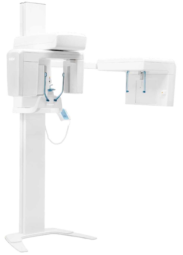
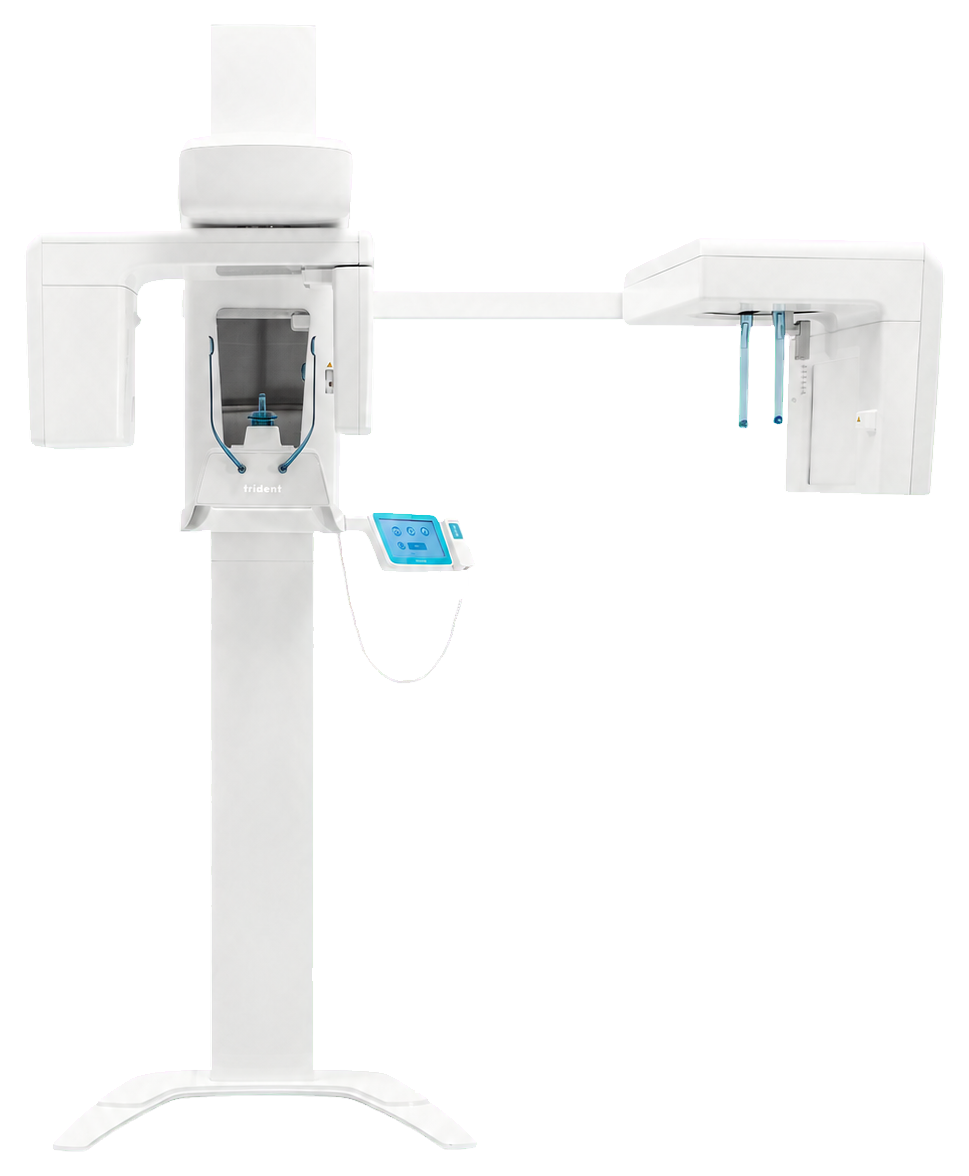
X‑VIEW 2D PAN CEPH is the complete 2‑in‑1 configuration, combining high-quality panoramic imaging and advanced cephalometric acquisition in one seamlessly integrated system.

Designed for demanding dental and orthodontic workflows, it enables practices to perform routine panoramic examinations, specialized projections and full-format cephalometric studies without changing equipment or interrupting the clinical workflow.

With two dedicated detectors—one optimized for panoramic imaging and one for cephalometric acquisition—each examination benefits from technology specifically designed for its purpose. The result is uncompromised performance, operational efficiency and exceptional versatility within a single platform.

DUAL-DETECTOR TECHNOLOGY

The high-resolution CMOS detector delivers detailed and consistent panoramic images, while the dedicated 25 × 30 cm Single‑Shot DR detector captures cephalometric projections in just 200–500 milliseconds.

Single‑Shot technology acquires the entire cephalometric image in one rapid exposure, helping minimize the effects of patient movement and ensuring a clear, geometrically consistent representation of the craniofacial structures.

DEEP‑VIEW + DEEP‑AI

X‑VIEW 2D PAN CEPH integrates seamlessly with Deep‑View, creating a smooth digital workflow from image acquisition to visualization and management.

When combined with the optional Deep‑AI module, artificial intelligence automatically enhances panoramic images by optimizing clarity, contrast, sharpness and image consistency—while preserving the original anatomical information.

Complete Capabilities. Seamless Workflow.
    • Panoramic and cephalometric imaging in one integrated system
    • Two dedicated detectors optimized for their respective applications
    • Full-format 25 × 30 cm LAT and PA cephalometric examinations
    • Single‑Shot cephalometric acquisition in just 200–500 ms
    • Adult panoramic examination in 15.5 seconds
    • Child panoramic examination in 14.8 seconds
    • Carpus imaging for skeletal maturity assessment
    • Variable field collimation
    • Unified software environment and patient database
    • Simple image storage, export and sharing

DIAGNOSTIC EXCELLENCE IN EVERY IMAGE

Every image acquired with X‑VIEW 2D PAN provides clear visualization of anatomical structures and clinically relevant details. Explore a wide selection of panoramic, cephalometric, and specialized examinations, and discover the image quality X‑VIEW 2D PAN brings to your daily practice.

2D PANORAMIC IMAGES

A single acquisition provides a comprehensive view of both dental arches, the jaws, and the surrounding structures, offering a complete basis for initial assessment and treatment planning.

CEPHALOMETRIC & ORTHODONTIC IMAGING

Cephalometric images provide a standardized representation of craniofacial structures, essential for assessing skeletal relationships, tooth position, and growth patterns. They support cephalometric analysis, orthodontic treatment planning, and progress monitoring over time.

OUR PRODUCTS

Effective products and solutions,
by professionals, for professionals.