X-VIEW 3D

Trident introduces the most advanced CBCT technology to acquire volumetric images with a single scan and less radiation dose.

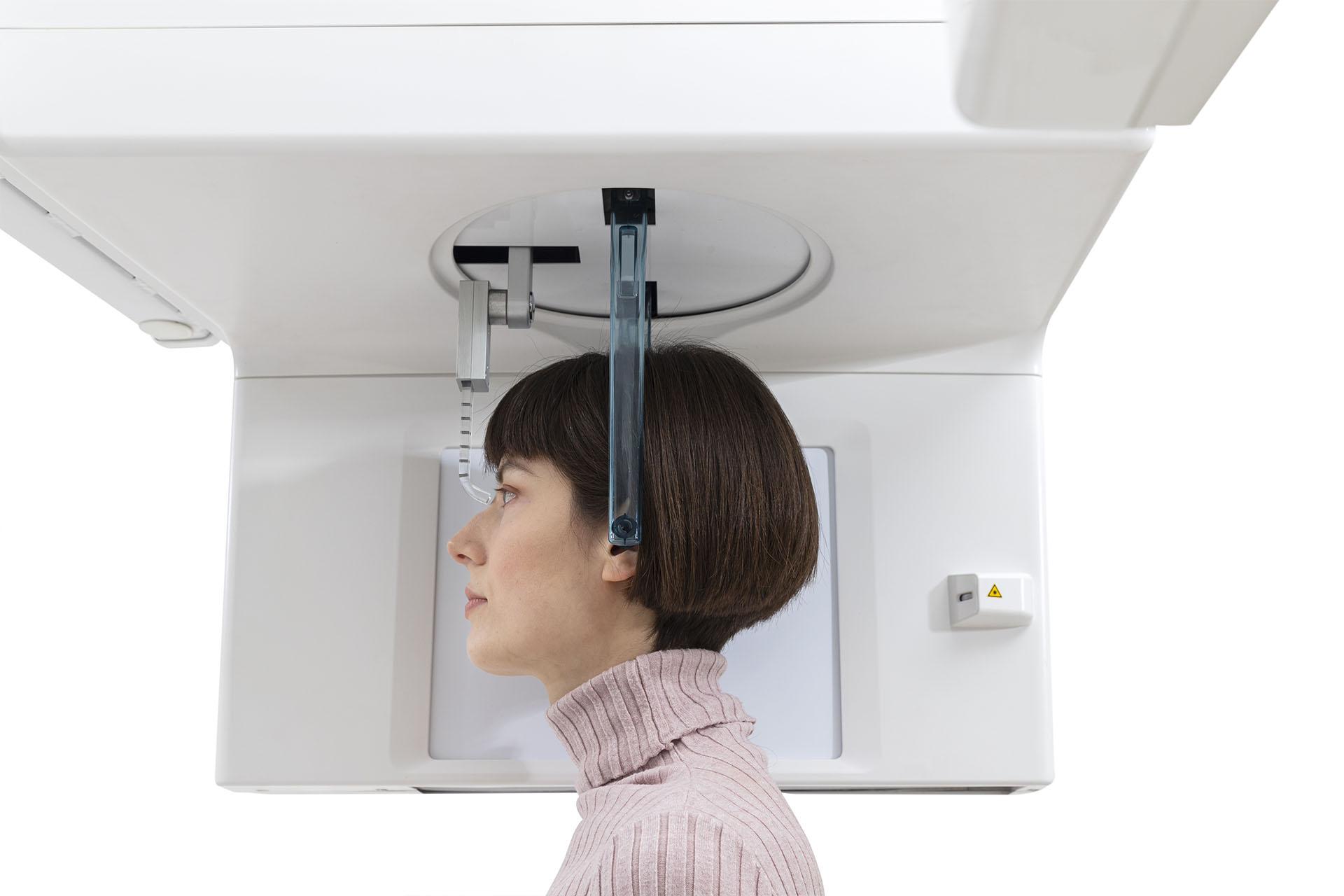
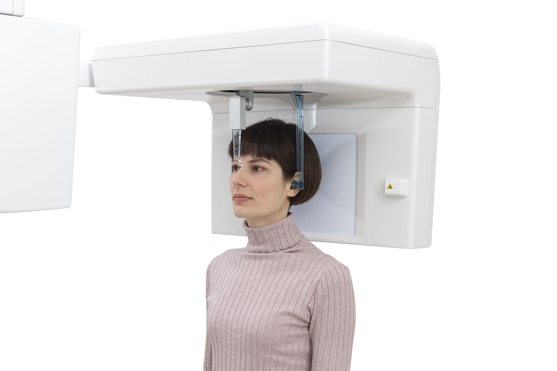
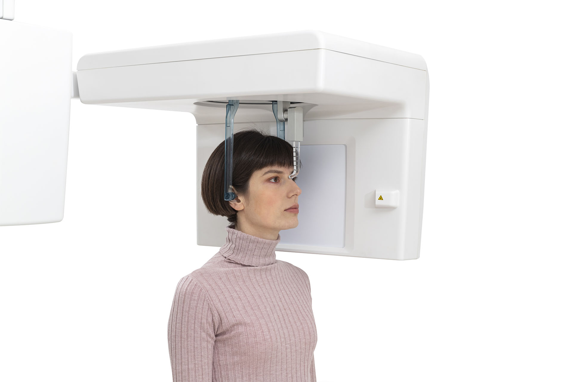
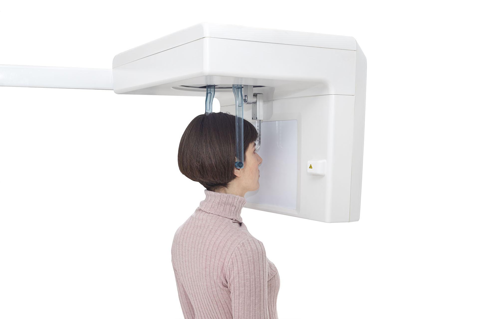
X-VIEW 3D system combines the latest advances in digital radiology with a clean and compact design to obtain high resolution 3D images of the dental structures, soft tissues, nerve paths and bone in the craniofacial region.


Available in three models:

X-VIEW 3D ONLYX-VIEW 3D PANX-VIEW 3D PAN CEPH
FEATURES

Remarkable features that
cannot be seen:

null
  • null
    PRECISE DIAGNOSTICS
    Images obtained with X-VIEW 3D cone beam CT allow for more precise diagnostics.
  • null
    ACCURATE IMAGES
    Thanks to its evolved functions, trajectories, and collimations applicable to each exam, images are full of details and contrast.
  • null
    HIGH CONNECTIVITY
    The software offers a wide range of tools to manage the images allowing to efficiently save, export and share files.
  • null
    LOW RADIATION DOSE
    The high frequency generator and the pulsed emission adjust the exposure adapting the dose to the dimensions of the examined area without compromising image quality.
  • null
    GO-GREEN AND SAFE PACKAGING
    Thanks to the new detachable column, the unit is safely packed using only one triple wall corrugated 120 * 80 * h120 cm cardboard box. It is a sustainable packaging manufactured with materials and techniques that reduce both energy consumption and the harmful impact of packaging on the environment.

Achieve flawless images with the innovative features integrated into X-VIEW 3D PAN, for outstanding results.

  • null
    3D CBCT RECONSTRUCTION
    Even in its Basic Version, without activating additional advanced features, the algorithm surpasses standard reconstruction techniques like filtered back projection (FBP) or Feldkamp (FDK).
  • null
    METAL ARTIFACT REDUCTION (MAR)
    Activate this option when high-density objects, like metallic components, are present within the scanned field. These objects typically lead to pronounced streaks and dark shadows.
  • null
    PATIENT MOTION CORRECTION
    Patient movement can lead to the appearance of double edges and blurred structures. This feature identifies and mitigates patient motion, ensuring sharp details and accurate reconstruction of anatomy.
  • null
    INCREASED HOMOGENEITY
    A dedicated reconstruction kernel tailored for dental scans enhances contrast, resulting in improved homogeneity and reduced brightening at the image periphery. The homogeneity factor determines the distribution of gray and/or color values within the image. Homogeneity is achieved when each pixel in the image shares the same color, while strong contrasts indicate inhomogeneity.
  • null
    ADVANCED CALIBRATION
    By using a specific geometric calibration tool and software module, it is possible to automatically and accurately calculate all of the scanner’s geometric parameters and compensate for any repeatable mechanical inaccuracies.

FOV STANDARD DOSE

X-VIEW 3D PAN offers multiple Field of View (FOV) options, while maintaining a standard dose for patient safety.
Experience precise imaging with customizable FOV settings that provide detailed visuals without compromising on radiation exposure.

FOV REDUCED DOSE

X-VIEW 3D PAN offers a larger field of view (FOV) for the same radiation dose, effectively reducing the dose per scan.
Benefit from wider image coverage without increasing radiation exposure, ensuring greater diagnostic accuracy with optimal patient safety.

X-VIEW 3D PAN allows image acquisition in three different resolution settings:

HD

Ideal for those seeking the highest quality in terms of high definition, although it requires more storage space.

STD

It represents an optimal compromise between image quality and file size.

WLF

A particularly lightweight version offering good image quality.

DOSEFOVVOXEL SIZE
cmRESOLUTION
STDHIGHWLF
STANDARD5×5655098
9×9176118352
6xØ11215143430
11×11215143430
REDUCED5xØ619550390
9xØ11176118352
6xØ14215143430
11xØ14215143430

Deep-View software for intraoral images.

Deep-View is a modern imaging software designed by Trident to efficiently acquire, organize, store and share digital images.
The suite is a complete and integrated software for intuitive navigation with several advanced features to obtain and manage thousands of high-definition images for more accurate diagnosis.

DFO: Dental - Facial and Orthodontics

Trident offers, as an optional, this functional tool for orthodontic tracing and cephalometric analysis.
DFO performs all kind of analysis in less time, just run the software and set the points needed for a complete analysis, the results will be automatically calculated and drawn on the screen.

Xelis - Dental implant planning software

Xelis is a professional tool to perform 3D implant simulation directly on the PC. The software allows the user to simulate the position of implants, identify the mandibular canal, trace panoramic views and sections of the bone model, view the three-dimensional bone model and calculate bone density. Doctors can easily and safely plan implant-prosthetic surgery.

The PC for X-VIEW 3D comes with Deep-View software + Xelis Basic software installed. The computer is configured from the factory in every single detail. The calibration files are already provided, so the user does not have to manually install them or even manipulate the computer with instructions for operation, avoiding complicated calibrations on site, after the installation, or during the unit useful life.

The ergonomic design ensures that the patient is not enforced to use awkward postures and can easily achieve the correct positioning.

The specially designed tools assist patients, during the acquisition process, to maintain their position avoiding errors in the procedures.

X-VIEW 3D PAN adapts to all sizes and types of patients, the linear and open design facilitates access for wheelchair users.

The LCD display enables users to directly interact with the unit.

Menus are very easy to navigate.

Universally recognized icons indicate available functions, parameters, and options.

All instructions are multilingual in a friendly language that guides the user fluently through the different options.

Available in three models to offer complete and affordable  imaging solutions:

X-VIEW 3D ONLY

X-VIEW 3D ONLY is perfect for doctors seeking simplicity and efficiency. With essential functions and straightforward operations, a single click is all it takes to obtain sharp and detailed 3D images.

IGZO-BASED FLAT-PANEL X-RAY DETECTOR
MINIMUM VOXEL SIZE 118 μm
SINGLE FOV 9x9 cm
SINGLE FOV 11x11 cm
SINGLE FOV 11xØ14 cm
DUAL FOV 11x11, 11xØ14 cm
EASY INSTALLATION AND MAINTENANCE
2D PANORAMIC IMAGE CAN BE RECONSTRUCTED FROM THE 3D EXAM BY SOFTWARE.

X-VIEW 3D ONLY provides the following display modes apart from basic orthogonal views:

OBLIQUE SLICING
CURVED SLICING
CROSS-SECTIONAL (OBLIQUE CORONAL) VIEW
ERAY SUM
VOLUME RENDERING

The intuitive collaboration of two software, Deep-View + Xelis, seamlessly operates on volumetric data. This includes tasks such as cutting, measuring bone density, assessing root depth, and analyzing the mandibular canal. This dynamic duo serves as an efficient tool for specialized procedures in endodontics, orthodontics, implantology, and maxillofacial surgery, empowering doctors with precise and specialized capabilities.

X-VIEW 3D ONLY offers a range of tools for analyzing and conducting implant treatments.

X-VIEW 3D PAN

X-VIEW 3D PAN is a 2-in-1 optimized solution that ensures a fast scanning experience (from 2.4 to 15.5 seconds) for a wide range of dental 2D panoramic and 3D images. Designed specifically for orthodontists, endodontists and implantologists, this unit is an effective tool for medium to large dental offices.

IGZO-BASED FLAT-PANEL X-RAY DETECTOR
PIXEL SIZE 100 μm
MINIMUM VOXEL SIZE 50 μm
SINGLE FOV 9x9 cm
SINGLE FOV 11x11 cm
SINGLE FOV 11xØ14 cm
DUAL FOV 11x11, 11xØ14 cm
MULTIFOV 5x5, 6xØ11, 9x9, 11x11 cm
MULTIFOV 5x5, 5xØ6, 6xØ11, 6xØ14, 9x9, 9xØ11, 11x11, 11xØ14 cm
15x30 cm ADULT 2D PAN IMAGES
13x30 cm PEDIATRIC 2D PAN IMAGES
VOLUMETRIC IMAGES
CEPH UPGRADABLE
  • Evaluates the detailed morphology of the bone tissue
  • Explores the maxillary sinuses
  • Detects dental anomalies
  • Determines the protocol for extraction of impacted teeth
  • Diagnoses temporomandibular joint problems

X-VIEW 3D PAN CEPH

This cutting-edge CBCT 3-in-1 system enables oral health professionals to conduct a wide range of examinations.
Alongside its outstanding 3D imaging capabilities, the X-VIEW 3D PAN CEPH features functionalities for 2D panoramic and cephalometric radiographs.

IGZO-BASED FLAT-PANEL X-RAY DETECTOR
PIXEL SIZE 100 μm
MINIMUM VOXEL SIZE 50 μm
VOLUMETRIC IMAGES
SINGLE FOV 9x9 cm
SINGLE FOV 11x11 cm
SINGLE FOV 11xØ14 cm
DUAL FOV 11x11, 11xØ14 cm
MULTIFOV 5x5, 6xØ11, 9x9, 11x11 cm
MULTIFOV 5x5, 5xØ6, 6xØ11, 6xØ14, 9x9, 9xØ11, 11x11, 11xØ14 cm
15x30 cm ADULT 2D PAN IMAGES
13x30 cm PEDIATRIC 2D PAN IMAGES
13x30 cm AMORPHOUS SILICON FLAT PANEL
30x25 cm AP AND LL CEPHALOGRAMS
CARPUS

AP and lateral cephalograms offer valuable insights for planning orthognathic surgery, correcting facial asymmetry, addressing malocclusion, and guiding orthodontic treatments.
The lateral cephalogram is utilized to determine the forward projection of nasion, subnasale, and gonion.
Meanwhile, the AP cephalogram assists in establishing the relationship between the horizontal occlusal plane and the skull base.

  • Surgical planning for impacted teeth
  • Diagnosis of TMJ disorder
  • Precise dental implant placement
  • Evaluation of the jaw, sinuses, nerve canals, and nasal cavity
  • Detect, measure and treat jaw tumors
  • Determination of bone structures and tooth orientation
  • Cephalometric analysis
OUR PRODUCTS

Effective products and solutions,
by professionals, for professionals.